Mpls. VA brain research examines resilience to trauma
New research from the Minneapolis VA Medical Center has identified brain patterns that appear to be markers of resilience to trauma. The findings could suggest why some people exposed to trauma develop post-traumatic stress disorder, or PTSD, while many others do not.
Researchers say the findings, published online Wednesday in the journal JAMA Psychiatry, point to a central mechanism showing how the brain can recover from traumatic events.
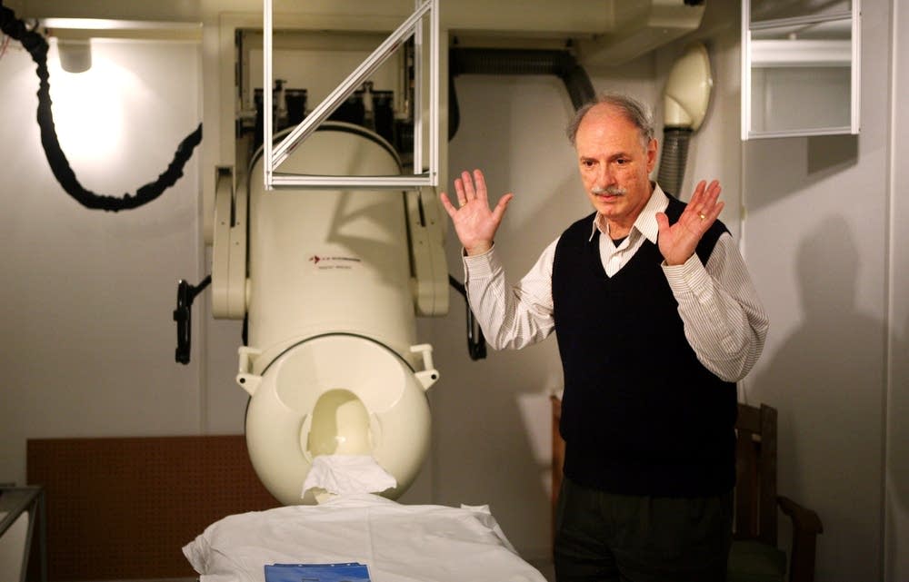
In the study, scientists compared the brains of nearly 200 veterans who had experienced trauma, using magnetoencephalography (MEG). The machine detects the magnetic fields produced when groups of brain cells communicate.
The MEG scans of PTSD-affected brains showed distinctive clusters of neurons locked into interactions with other clusters.
Create a More Connected Minnesota
MPR News is your trusted resource for the news you need. With your support, MPR News brings accessible, courageous journalism and authentic conversation to everyone - free of paywalls and barriers. Your gift makes a difference.
"We believe these neuron networks were stuck in the trauma-encoding phase," said lead author Lisa James, a research psychologist at the Brain Sciences Center and an assistant psychiatry professor at the University of Minnesota. "The trauma had a hold on them. They weren't available to encode new information."
She compared the phenomenon to a "phone network where every line is busy."
By contrast, James said, non-PTSD volunteers showed no such patterns.
Their neural networks were flexible, adaptable and available. They were free to link up with other neuron groups as needed to react to new incoming experiences.
"People really are resilient," James said, "and we've been able to demonstrate exactly what is going on in the brain that helps facilitate healthy functioning following trauma exposure."
The study's co-author, Brian Engdahl, a psychologist at the Minneapolis VA and an associate psychology professor at the University of Minnesota, said PTSD-affected brains look very different from normal brains, which process trauma and move on to other tasks.
"Those who have continued to experience disorders such as PTSD have not yet adapted their brain function to accommodate the trauma," Engdahl said, "and we are actively investigating the best treatment routes to produce those changes."
Engdahl used the analogy of a handshake to describe how healthy neural networks respond to traumas: "When a trauma comes in, everyone [populations of neurons within the brain] is shaking hands tightly. The neurons are very tied up with processing this experience. Over time, the handshake weakens. It gets less intense. The network eventually gets released and is free to respond to other events."
In normal brains, he said, the traumatic memory eventually gets consolidated and deposited in other brain regions. The memory remains housed in the brain, but it's not pathological. It does not impair function.
The brain area where the researchers saw the sharpest difference between the two groups of study volunteers was the right superior temporal gyrus.
Part of this region in the brain helps with auditory processing, but the researchers say the area they were probing was a deeper part of the superior temporal gyrus, the role of which is not clearly known.
"It's no man's land," said senior author Dr. Apostolos Georgopoulos, director of the Brain Sciences Center at the Minneapolis VA and Regents Professor in the University of Minnesota Medical School's Department of Neuroscience.
He pointed out, though, that some studies have linked the region to the re-experiencing of past events, which clearly plays a role in PTSD.
"Our patients with PTSD tell us that the intrusive memories have a life of their own," Georgopoulos said. "They pop into their minds when they don't want them. And that's what we're seeing on the brain scans."
The new study complements research published by the group in 2010 in which it first described distinctive PTSD brain patterns, as detected by the MEG.
"This new paper helps explain why we found what we found in those earlier studies," Georgopoulos said. "Based on this work, we hope to develop reliable biomarkers for PTSD and for emotional resilience."
The study was partly supported by a grant from the Department of Veterans Affairs.
See a copy of the original JAMA article below.
